58 minutes ago
2
58 minutes ago
2

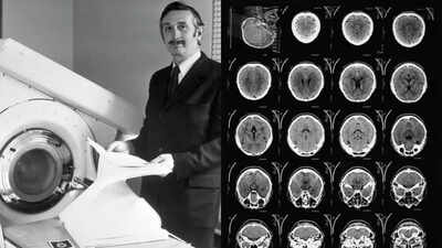
Godfrey Hounsfield stands beside the EMI-Scanner in 1972 and modern CT scans provide much higher resolution images of the 'slices' of the brain than Hounsfield's original scan did in 1971.
What if the contents of a sealed box could be revealed without ever opening it? For British engineer Godfrey Hounsfield, the question began with an unlikely fascination with Egyptian pyramids and cosmic rays.
Years later, that same idea would lead to a machine capable of looking through the skull and revealing the soft tissue of the human brain. On October 1, 1971, his invention produced the first clinical CT image of a human brain, identifying a cyst that conventional X-rays could not clearly show, states an online report by Smithsonian Magazine.
From radar to an extraordinary idea
According to the report, Hounsfield's career began far from medicine. During the Second World War, he worked with radar in the Royal Air Force, where his talent for electrical equipment quickly became apparent.
After the war, he trained as an engineer and joined EMI, the British electronics company that would later become famous for its association with The Beatles. By the 1960s, Hounsfield had become a leading figure in computing.
When EMI withdrew from the computer business, however, he found himself with greater freedom to pursue unconventional ideas.One of them involved seeing inside objects without opening them.
His interest in whether cosmic rays could reveal hidden spaces inside pyramids eventually evolved into a medical question after he encountered the limitations of conventional brain X-rays. Bones appeared clearly, but soft brain tissue largely merged into an indistinct mass.
Turning the brain into slices
Hounsfield's solution was deceptively simple in concept. He imagined dividing the brain into a series of thin slices, like cutting a loaf of bread.
X-rays could then be directed through each layer from numerous angles, with detectors recording how much radiation passed through, adds the online report.Different tissues absorb X-rays differently. By collecting thousands of measurements and feeding them into a computer, Hounsfield developed a method to calculate the density of tiny sections within each slice and reconstruct them into an image, the report adds.If we go by the report, the first machines were far from today's sleek scanners. Working with limited resources, Hounsfield assembled an experimental system from equipment and components available around EMI's research facilities. Early tests on objects and animal brains demonstrated that the concept could distinguish structures that ordinary X-rays could not.
From laboratory experiment to hospital
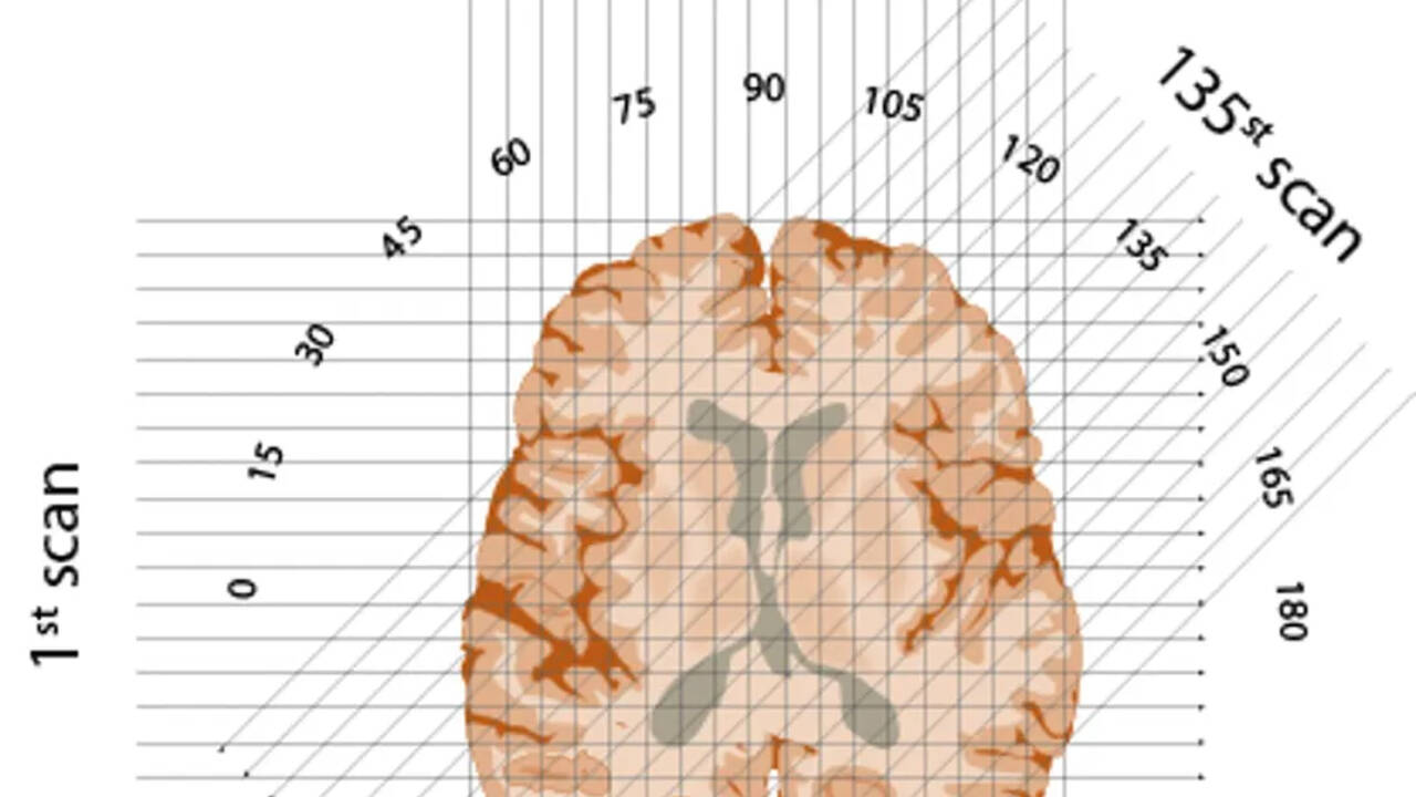
X-rays beam through each ‘slice’ of brain, oriented at each degree from 1 to 180 in a semicircle
The biggest obstacle was proving the technology could work safely on people.
Hounsfield's project received crucial support from within EMI and eventually from Britain's Department of Health and Social Security, which agreed to purchase scanners for hospitals even before the machines were completed.A full-sized scanner was installed at Atkinson Morley Hospital in London. On October 1, 1971, a woman with suspected brain disease became the first patient to undergo the procedure.The process was painfully slow by modern standards. The scan took about 30 minutes, while the data had to be transported across London on magnetic tapes for processing by an EMI mainframe.
Several hours later, an image could finally be produced, explains the online report from 2021. The result was unmistakable: a cystic mass was visible in the patient's frontal lobe, it adds.
A medical revolution
The breakthrough quickly changed the way doctors investigated disease inside the body. CT imaging made it possible to locate tumours, bleeding and other abnormalities without exploratory surgery.The technology soon expanded beyond the brain. In 1973, American engineer Robert Ledley developed a whole-body CT scanner, opening the door to imaging organs, blood vessels and other structures.Hounsfield shared the 1979 Nobel Prize in Physiology or Medicine for the development of computer-assisted tomography. He was knighted in 1981 and continued working on inventions until his death in 2004. Today, CT scanners are vastly faster, sharper and more efficient, with substantially lower radiation exposure than early machines. More than 80 million CT scans were being performed annually in the United States by 2020, illustrating just how deeply Hounsfield's idea became embedded in modern medicine.And his original fascination with hidden spaces came full circle. Cosmic-ray detectors have since been used to investigate the pyramids, including the discovery of a previously unknown chamber inside the Great Pyramid of Giza in 2017. The engineer who wanted to see inside a pyramid ultimately gave medicine a way to see inside us.Images Courtesy: Smithsonian Magazine